Maternal Free Fatty Acids and Newborn Hypothalamus
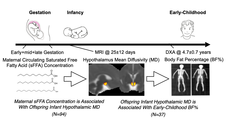
This study tested the hypothesis, in a prospective cohort study design, that maternal saturated free fatty acid (sFFA) concentration during pregnancy is prospectively associated with offspring (newborn) hypothalamic (HTH) microstructure and to explore the functional relevance of this association with respect to early-childhood body fat percentage (BF%). In N = 94 healthy newborns (born mean 39.3 [SD 1.5] weeks gestation), diffusion-weighted magnetic resonance imaging was performed shortly after birth (25.3 [12.5] postnatal days), and a subgroup (n = 37) underwent a dual-energy x-ray absorptiometry scan in early childhood (4.7 [SD 0.7] years). Maternal sFFA concentration during pregnancy was quantified in fasting blood samples via liquid chromatography-mass spectrometry. Infant HTH microstructural integrity was characterized using mean diffusivity (MD). Multiple linear regression was used to test the association between maternal sFFA and HTH MD, accounting for newborn sex, age at scan, mean white matter MD, and image quality. Multiple linear regression models also tested the association between HTH MD and early-childhood BF%, accounting for breastfeeding status. Maternal sFFA during pregnancy accounted for 8.3% of the variation in newborn HTH MD (β-std = 0.25; p = 0.006). Furthermore, newborn HTH MD prospectively accounted for 15% of the variation in early-childhood BF% (β-std = 0.32; p = 0.019). These findings suggest that maternal overnutrition during pregnancy may influence the development of the fetal hypothalamus, which, in turn, may have clinical relevance for childhood obesity risk.
Maternal IL-6 and Childhood Fluid Intelligence
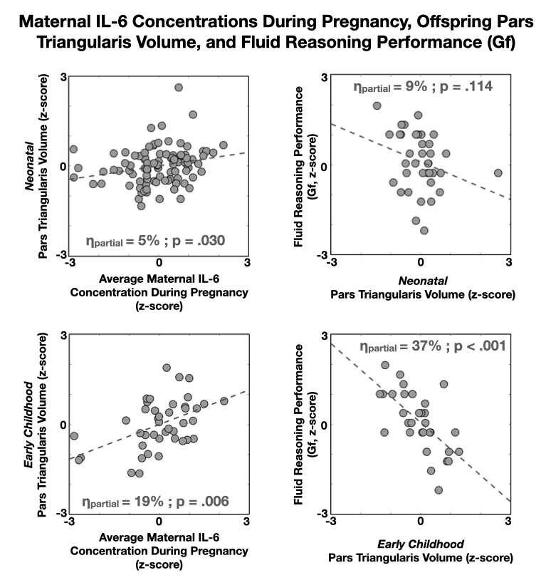
Maternal inflammation during pregnancy can alter offspring brain development and influence risk for disorders commonly accompanied by deficits in cognitive functioning. We therefore examined associations between maternal interleukin 6 (IL-6) concentrations during pregnancy and offspring cognitive ability and concurrent magnetic resonance imaging-based measures of brain anatomy in early childhood. We further examined newborn brain anatomy in secondary analyses to consider whether effects are evident soon after birth and to increase capacity to differentiate effects of pre- versus postnatal exposures. IL-6 concentrations were quantified in early (12.6 ± 2.8 weeks), mid (20.4 ± 1.5 weeks), and late (30.3 ± 1.3 weeks) pregnancy. Offspring nonverbal fluid intelligence (Gf) was assessed at 5.2 ± 0.6 years using a spatial reasoning task (Wechsler Preschool and Primary Scale of Intelligence-Matrix) (n = 49). T1-weighted magnetic resonance imaging scans were acquired at birth (n = 89, postmenstrual age = 42.9 ± 2.0 weeks) and in early childhood (n = 42, scan age = 5.1 ± 1.0 years). Regional cortical volumes were examined for a joint association between maternal IL-6 and offspring Gf performance. Average maternal IL-6 concentration during pregnancy was inversely associated with offspring Gf performance after adjusting for socioeconomic status and the quality of the caregiving and learning environment (R2 = 13%; p = .02). Early-childhood pars triangularis volume was jointly associated with maternal IL-6 and childhood Gf (pcorrected < .001). An association also was observed between maternal IL-6 and newborn pars triangularis volume (R2 = 6%; p = .02). These findings suggest that the origins of variation in child cognitive ability can, in part, trace back to maternal conditions during the intrauterine period of life and support the role of inflammation as an important component of this putative biological pathway.
Maternal IL-6 and Newborn Uncinate Fasciculus
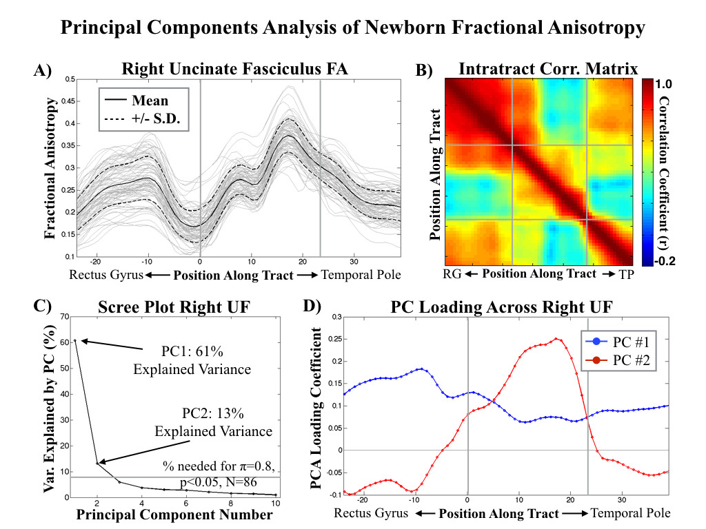
Maternal inflammation during pregnancy can alter the trajectory of fetal brain development and increase risk for offspring psychiatric disorders. However, the majority of relevant research to date has been conducted in animal models. Here, in humans, we focus on the structural connectivity of frontolimbic circuitry as it is both critical for socioemotional and cognitive development, and commonly altered in a range of psychiatric disorders associated with intrauterine inflammation. Specifically, we test the hypothesis that elevated maternal concentration of the proinflammatory cytokine interleukin-6 (IL-6) during pregnancy will be associated with variation in microstructural properties of this circuitry in the neonatal period and across the first year of life. Pregnant mothers were recruited in early pregnancy and maternal blood samples were obtained for assessment of maternal IL-6 concentrations in early (12.6 ± 2.8 weeks [S.D.]), mid (20.4 ± 1.5 weeks [S.D.]) and late (30.3 ± 1.3 weeks [S.D.]) gestation. Offspring brain MRI scans were acquired shortly after birth (N = 86, scan age = 3.7 ± 1.7 weeks [S.D.]) and again at 12-mo age (N = 32, scan age = 54.0 ± 3.1 weeks [S.D.]). Diffusion Tensor Imaging (DTI) was used to characterize fractional anisotropy (FA) along the left and right uncinate fasciculus (UF), representing the main frontolimbic fiber tract. In N = 30 of the infants with serial MRI data at birth and 12-mo age, cognitive and socioemotional developmental status was characterized using the Bayley Scales of Infant Development. All analyses tested for potentially confounding influences of household income, prepregnancy Body-Mass-Index, obstetric risk, smoking during pregnancy, and infant sex, and outcomes at 12-mo age were additionally adjusted for the quality of the postnatal caregiving environment. Maternal IL-6 concentration (averaged across pregnancy) was prospectively and inversely associated with FA (suggestive of reduced integrity under high inflammatory conditions) in the newborn offspring (bi-lateral, p < 0.01) in the central portion of the UF proximal to the amygdala. Furthermore, maternal IL-6 concentration was positively associated with rate of FA increase across the first year of life (bi-lateral, p < 0.05), resulting in a null association between maternal IL-6 and UF FA at 12-mo age. Maternal IL-6 was also inversely associated with offspring cognition at 12-mo age, and this association was mediated by FA growth across the first year of postnatal life. Findings from the current study support the premise that susceptibility for cognitive impairment and potentially psychiatric disorders may be affected in utero, and that maternal inflammation may constitute an intrauterine condition of particular importance in this context.
Maturation Index
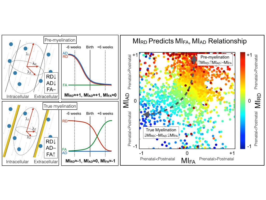
Human birth presents an abrupt transition from intrauterine to extrauterine life. Here we introduce a novel Maturation Index (MI) that considers the relative importance of gestational age at birth and postnatal age at scan in a General Linear Model. The MI is then applied to Diffusion Tensor Imaging (DTI) in newborns for characterizing typical white matter development in neonates. DTI was performed cross-sectionally in 47 neonates (gestational age at birth=39.1±1.6 weeks [GA], postnatal age at scan=25.5±12.2 days [SA]). Radial diffusivity (RD), axial diffusivity (AD) and fractional anisotropy (FA) along 27 white matter fiber tracts were considered. The MI was used to characterize inflection in maturation at the time of birth using GLM estimated rates of change before and after birth. It is proposed that the sign (positive versus negative) of MI reflects the period of greatest maturation rate. Two general patterns emerged from the MI analysis. First, RD and AD (but not FA) had positive MI on average across the whole brain (average MIAD=0.31+/−.42, average MIRD=0.22+/−.34). Second, significant regions of negative MI in RD and FA (but not AD) were observed in the inferior corticospinal regions, areas known to myelinate early. Observations using the proposed method are consistent with proposed models of the white matter maturation process in which pre-myelination is described by changes in AD and RD due to oligodendrocyte proliferation while true myelination is characterized by changes in RD and FA due to myelin formation.
Marijuana Use and ADHD in Adolescents
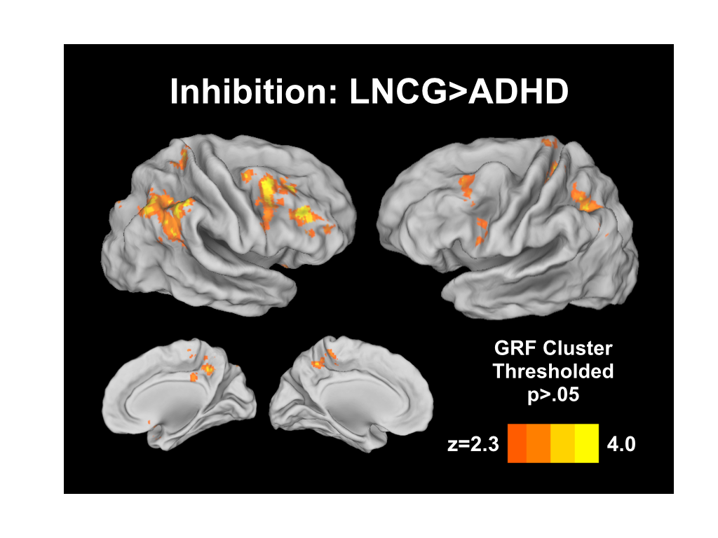
Children diagnosed with attention-deficit/hyperactivity disorder (ADHD) are at increased risk for substance abuse. Response inhibition is a hallmark of ADHD, yet the combined effects of ADHD and regular substance use on neural networks associated with response inhibition are unknown. Task-based functional Magnetic Resonance Imaging (fMRI) data from young adults with childhood ADHD with (n = 25) and without (n = 25) cannabis use ≥ monthly in the past year were compared with a local normative comparison group (LNCG) with (n = 11) and without (n = 12) cannabis use. Go/NoGo behavioral and fMRI data were evaluated for main and interaction effects of ADHD diagnosis and cannabis use. ADHD participants made significantly more commission errors on NoGo trials than controls. ADHD participants also had less frontoparietal and frontostriatal activity, independent of cannabis use. No main effects of cannabis use on response inhibition or functional brain activation were observed. An interaction of ADHD diagnosis and cannabis use was found in the right hippocampus and cerebellar vermis, with increased recruitment of these regions in cannabis-using controls during correct response inhibition. ADHD participants had impaired response inhibition combined with less fronto-parietal/striatal activity, regardless of cannabis use history. Cannabis use did not impact behavioral response inhibition. Cannabis use was associated with hippocampal and cerebellar activation, areas rich in cannabinoid receptors, in LNCG but not ADHD participants. This may reflect recruitment of compensatory circuitry in cannabis using controls but not ADHD participants. Future studies targeting hippocampal and cerebellar-dependent function in these groups may provide further insight into how this circuitry is altered by ADHD and cannabis use.